
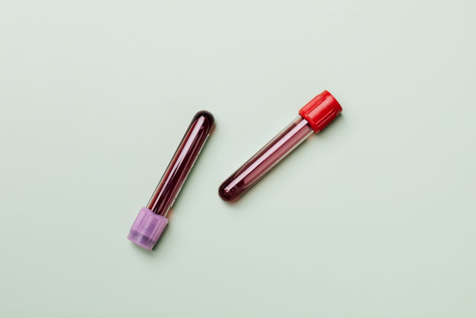
Red marrow is the soft, spongy tissue inside your bones that manufactures virtually every blood cell circulating in your body — roughly 200 billion red blood cells, 10 billion white blood cells, and 400 billion platelets every single day. When red marrow functions properly, you never think about it. When it doesn’t, the consequences range from chronic fatigue to life-threatening blood cancers. Unraveling the role of red marrow in health and disease starts with appreciating just how much your body depends on this often-overlooked tissue.
If you’re reading this because a doctor mentioned your bone marrow on imaging or lab work flagged something unusual, here’s the bottom line: red marrow is where blood is born. Problems with it show up as abnormal blood counts — low hemoglobin, weird white cell numbers, or platelets that are too high or too low. A complete blood count (CBC) is usually the first screening tool, and a bone marrow biopsy is the gold standard for direct evaluation.
What Exactly Does Red Marrow Do?
Red marrow’s primary job is hematopoiesis — the production of all three major blood cell lineages from a single pool of hematopoietic stem cells (HSCs). These pluripotent stem cells sit at the top of a differentiation hierarchy, branching into myeloid and lymphoid progenitors that ultimately become mature, functional cells.
In adults, active red marrow is concentrated in the axial skeleton: the pelvis (which contains about 40% of total red marrow), vertebrae, sternum, ribs, skull, and the proximal ends of the femur and humerus. By contrast, the shafts of long bones in adults contain mostly yellow marrow — essentially fat storage.
Red Marrow vs. Yellow Marrow: Key Differences
| Feature | Red Marrow | Yellow Marrow |
|---|---|---|
| Primary composition | Hematopoietic stem cells, developing blood cells | Adipocytes (fat cells) |
| Main function | Blood cell production | Fat/energy storage |
| Location in adults | Pelvis, vertebrae, sternum, ribs, skull, proximal femur/humerus | Shafts of long bones (tibia, fibula, distal femur) |
| MRI signal (T1-weighted) | Low to intermediate signal | High signal (bright, like subcutaneous fat) |
| Changes with age | Decreases; converts to yellow marrow from childhood onward | Increases with age |
| Reconversion potential | N/A | Can revert to red marrow under physiologic stress |
This conversion process is clinically relevant. At birth, nearly all bone marrow is red. By age 25, roughly 50% has converted to yellow marrow, and that ratio continues shifting. However, during severe anemia, chronic hypoxia, or heavy blood loss, yellow marrow can reconvert back to red marrow — a finding sometimes seen incidentally on MRI and occasionally mistaken for pathology.
Diseases That Directly Affect Red Marrow
When red marrow fails or malfunctions, the clinical fallout is significant. Here are the major categories:
Bone Marrow Failure Syndromes
- Aplastic anemia — The marrow stops producing enough cells across all three lineages (pancytopenia). Severe aplastic anemia has a mortality rate exceeding 70% within two years if untreated. Annual incidence is about 2–3 per million in Western countries.
- Myelodysplastic syndromes (MDS) — The marrow produces blood cells, but they’re defective and die prematurely. MDS affects roughly 4 per 100,000 people annually, with incidence rising sharply after age 60. About one-third of MDS cases progress to acute myeloid leukemia.
Myeloproliferative Neoplasms
- Polycythemia vera — Overproduction of red blood cells, often driven by a JAK2 V617F mutation (present in ~95% of cases).
- Essential thrombocythemia — Excess platelet production, raising clotting risk.
- Primary myelofibrosis — Progressive scarring of the marrow, forcing blood production to shift to the spleen and liver (extramedullary hematopoiesis).
Blood Cancers Involving Red Marrow
- Leukemia — Malignant proliferation of abnormal white blood cells within the marrow. Acute forms (AML, ALL) can fill the marrow so completely that normal blood cell production collapses within weeks.
- Multiple myeloma — Cancerous plasma cells accumulate in the marrow, causing bone destruction, anemia, and kidney damage. Median age at diagnosis is 69.
- Lymphoma — Can infiltrate bone marrow in advanced stages, particularly in non-Hodgkin lymphoma (found in marrow in 30–50% of cases at staging).
Metastatic Disease
Solid tumors — especially breast, prostate, lung, kidney, and thyroid cancers — frequently metastasize to red marrow. The pelvis and spine are the most common sites, which makes sense given their rich blood supply and high red marrow content.
How Doctors Evaluate Red Marrow
Evaluation typically follows a stepwise approach:
- CBC with differential — The first-line screening test. Abnormalities in hemoglobin (normal: 12–16 g/dL in women, 14–18 g/dL in men), WBC count (4,500–11,000/µL), or platelets (150,000–400,000/µL) can signal marrow dysfunction.
- Peripheral blood smear — Microscopic examination of blood cell shape and maturity. Blasts, teardrop cells, or hypersegmented neutrophils each point toward specific marrow pathologies.
- Bone marrow biopsy and aspirate — A needle is inserted into the posterior iliac crest (back of the pelvis) to obtain a core of marrow tissue. This provides cellularity data — normal adult marrow cellularity is roughly “100 minus age” percent (so a 60-year-old should have approximately 40% cellular marrow).
- MRI — Increasingly used to assess marrow composition non-invasively. Red marrow reconversion, infiltrative disease, and marrow edema are all identifiable on MRI without a biopsy.
When to See a Doctor
You should seek medical evaluation if you experience:
- Persistent, unexplained fatigue that doesn’t improve with rest
- Frequent or unusual infections
- Easy bruising or bleeding that seems disproportionate to injury
- Unexplained bone pain, particularly in the spine, pelvis, or ribs
- A CBC showing any cytopenia (low counts) or unexplained elevations
Ask your doctor specifically: “Could this be a bone marrow issue, and do I need a hematology referral?” Primary care physicians can screen with a CBC, but a hematologist is the specialist who interprets marrow biopsies and manages marrow disorders.
Frequently Asked Questions
Can red marrow regenerate after damage?
Yes, to a degree. After chemotherapy, red marrow typically recovers within 2–4 weeks, depending on the regimen. After a bone marrow transplant, engraftment — the point where new stem cells begin producing blood cells — usually occurs around day 14–21. However, some insults (like high-dose radiation) can permanently destroy marrow if stem cells are wiped out completely.
Why does red marrow show up on my MRI report?
Radiologists note red marrow because its appearance can mimic disease. Red marrow reconversion — where yellow marrow converts back to red — happens in smokers, obese individuals, long-distance runners, people living at high altitude, and anyone with chronic anemia. It’s usually benign but needs to be distinguished from marrow infiltration by cancer.
Does diet affect red marrow function?
Absolutely. Red marrow needs iron, vitamin B12, folate, and copper to produce healthy blood cells. Iron deficiency is the most common nutritional cause of impaired erythropoiesis worldwide, affecting an estimated 1.2 billion people. Severe B12 or folate deficiency causes megaloblastic changes visible on marrow biopsy.
What’s the difference between a bone marrow biopsy and an aspirate?
They’re usually done together but provide different information. The aspirate suctions out liquid marrow for microscopic evaluation of individual cell morphology and for flow cytometry. The biopsy removes a solid core of tissue (typically 1.5–2 cm) to assess overall architecture, cellularity, and fibrosis. Both are obtained from the same site in a single procedure that takes about 15–20 minutes.
At what age does red marrow stop being active?
Red marrow never fully stops — it just shrinks in distribution. The conversion from red to yellow marrow begins in childhood, starting in the fingers and toes, and progresses centrally. By age 25, red marrow is largely confined to the axial skeleton and proximal long bones, where it remains active for life.


