
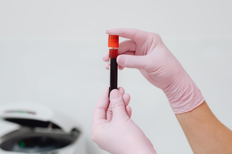
Haematologic malignancies are cancers that arise in the blood, bone marrow, or lymphatic system. They include leukemia, lymphoma, myeloma, and myelodysplastic syndromes — and collectively, they account for roughly 10% of all new cancer diagnoses worldwide, with over 180,000 new cases per year in the United States alone. If you’re looking for a comprehensive overview of haematologic malignancies, this guide covers the major types, how they’re classified, what symptoms to watch for, and what modern treatment looks like.
The term “haematologic malignancy” is an umbrella — beneath it sit dozens of distinct diseases, each with different biology, prognosis, and treatment strategies. A 5-year-old with acute lymphoblastic leukemia and a 72-year-old with chronic lymphocytic leukemia have dramatically different diseases, even though both fall under “blood cancer.” That distinction matters enormously for prognosis and treatment decisions.
The Major Categories of Haematologic Malignancies
Blood cancers are broadly divided into three main families, with several important subcategories within each.
Leukemias
Leukemias originate in the bone marrow and flood the bloodstream with abnormal white blood cells. They’re classified by speed of progression (acute vs. chronic) and by cell lineage (lymphoid vs. myeloid), giving us four core types: ALL, AML, CLL, and CML.
Lymphomas
Lymphomas arise in the lymphatic system — lymph nodes, spleen, thymus, and associated tissues. The two main categories are Hodgkin lymphoma (HL), which is characterized by Reed-Sternberg cells, and non-Hodgkin lymphoma (NHL), which encompasses over 60 distinct subtypes ranging from indolent (slow-growing) to highly aggressive.
Plasma Cell Neoplasms
Multiple myeloma is the most common plasma cell malignancy. It causes abnormal plasma cells to accumulate in the bone marrow, producing a dysfunctional antibody (M-protein) and often leading to bone destruction, kidney damage, and immunodeficiency.
Haematologic Malignancies at a Glance: Comparison Table
| Type | Median Age at Diagnosis | 5-Year Survival Rate | Key Feature |
|---|---|---|---|
| ALL (Acute Lymphoblastic Leukemia) | ~15 years | ~90% (children); ~40% (adults) | Most common childhood cancer |
| AML (Acute Myeloid Leukemia) | ~68 years | ~30% overall | Rapid onset; Auer rods on smear |
| CLL (Chronic Lymphocytic Leukemia) | ~72 years | ~87% | Often watch-and-wait initially |
| CML (Chronic Myeloid Leukemia) | ~64 years | ~70% | Philadelphia chromosome (BCR-ABL1) |
| Hodgkin Lymphoma | ~39 years | ~89% | Bimodal age distribution |
| Non-Hodgkin Lymphoma | ~67 years | ~74% | 60+ subtypes; highly variable |
| Multiple Myeloma | ~69 years | ~58% | Bone lesions, M-protein, renal failure |
Survival data from SEER Cancer Statistics Review. Individual prognosis varies significantly based on molecular subtype, stage, and patient fitness.
Symptoms That Should Raise a Red Flag
Haematologic malignancies are notorious for presenting with vague, nonspecific symptoms — which is exactly why they’re often diagnosed late. Here’s what to watch for:
- Persistent fatigue that doesn’t improve with rest (often from anemia — hemoglobin below 10 g/dL)
- Recurrent or unusual infections — a sign of neutropenia or dysfunctional immune cells
- Easy bruising or bleeding — petechiae, gum bleeding, or heavy periods from low platelets (below 100,000/µL)
- Drenching night sweats that soak through clothing
- Unexplained weight loss of more than 10% of body weight over 6 months
- Painless, progressively enlarging lymph nodes — especially if they persist beyond 4–6 weeks
- Bone pain — particularly in the back or ribs, classic for myeloma
- Persistent fever without an obvious infectious source
The triad of night sweats, weight loss, and fever — known as “B symptoms” in lymphoma staging — carries prognostic significance and should always prompt further workup.
Causes and Risk Factors
For most patients, there’s no single identifiable cause. Haematologic malignancies result from acquired genetic mutations in blood-forming stem cells or immune cells. But certain factors clearly increase risk:
- Age: The single biggest risk factor for most subtypes. AML, CLL, myeloma, and NHL all rise sharply after age 60.
- Prior chemotherapy or radiation: Alkylating agents and topoisomerase II inhibitors are linked to therapy-related AML/MDS, typically appearing 2–10 years after treatment.
- Chemical exposures: Benzene (occupational exposure) is a well-established risk factor for AML.
- Viral infections: EBV is linked to Burkitt lymphoma and some Hodgkin lymphoma cases. HTLV-1 causes adult T-cell leukemia/lymphoma. HIV increases NHL risk by 60–200 fold.
- Genetic syndromes: Down syndrome carries a 10–20× increased risk of childhood leukemia. Li-Fraumeni, Fanconi anemia, and ataxia-telangiectasia also raise risk.
- Family history: First-degree relatives of CLL patients have a 6–9× higher risk of developing CLL themselves.
How Haematologic Malignancies Are Diagnosed
Diagnosis typically starts with blood work and escalates based on findings. Here’s the standard workup:
Initial Laboratory Tests
- Complete blood count (CBC) with differential: Reveals cytopenias (low counts) or leukocytosis (elevated WBC). A WBC above 100,000/µL is a medical emergency (leukostasis).
- Peripheral blood smear: Manual review by a pathologist can identify blasts, atypical lymphocytes, or rouleaux formation (myeloma).
- Comprehensive metabolic panel: Elevated LDH, calcium, creatinine, and uric acid can suggest tumor burden or organ damage.
- Serum protein electrophoresis (SPEP): Essential for detecting the monoclonal protein in myeloma.
Bone Marrow Biopsy
This remains the gold standard for most haematologic malignancies. A sample is taken from the posterior iliac crest and analyzed for cellularity, blast percentage (≥20% defines acute leukemia by WHO criteria), and architectural changes. Flow cytometry, cytogenetics, and molecular testing (FISH, NGS) are performed simultaneously to classify the disease precisely.
Imaging
PET/CT is the standard for staging lymphoma and assessing treatment response. CT and MRI are used for evaluating organomegaly, bone lesions (myeloma), and CNS involvement. Whole-body low-dose CT has largely replaced skeletal surveys for myeloma bone disease.
Modern Treatment Approaches
Treatment for haematologic malignancies has evolved dramatically over the past two decades. Here’s a snapshot of the current landscape:
- Chemotherapy: Still the backbone for acute leukemias and aggressive lymphomas, but increasingly combined with targeted agents.
- Targeted therapy: Tyrosine kinase inhibitors (imatinib, dasatinib) transformed CML from a fatal disease to one with near-normal life expectancy. BTK inhibitors (ibrutinib, acalabrutinib) have reshaped CLL treatment.
- Immunotherapy: Monoclonal antibodies (rituximab for B-cell lymphomas), bispecific antibodies (blinatumomab), and immune checkpoint inhibitors (nivolumab for relapsed Hodgkin lymphoma) are now standard of care.
- CAR-T cell therapy: Approved for relapsed/refractory ALL, DLBCL, and multiple myeloma. Complete response rates of 50–80% in heavily pretreated patients.
- Stem cell transplantation: Allogeneic transplant remains the only curative option for many high-risk leukemias. Autologous transplant is standard consolidation for eligible myeloma patients.
When to See a Doctor
Get evaluated promptly if you experience any of the following:
- Persistent fatigue, pallor, or shortness of breath that’s new or worsening
- Unexplained bruising, petechiae, or bleeding that’s out of proportion to injury
- A lymph node that’s been swollen for more than 4 weeks and is painless, firm, and growing
- Recurrent infections — especially if you’re getting sick more often or more severely than usual
- Drenching night sweats or unexplained weight loss exceeding 10% over 6 months
A simple CBC can identify many of these cancers — or at least raise a flag that warrants referral to a hematologist. Don’t wait for symptoms to become severe.
Key Takeaways
- Haematologic malignancies represent a diverse group of over 100 distinct diseases — not a single condition.
- They account for ~10% of cancers and over 180,000 new US cases per year.
- Survival has improved dramatically: 5-year survival for Hodgkin lymphoma exceeds 89%, and CML patients on TKIs have near-normal life expectancy.
- Early detection through CBC and timely referral to a hematologist remain the most impactful steps you can take.
- Molecular profiling (cytogenetics, NGS) now drives treatment decisions as much as the disease name itself.
Frequently Asked Questions
Are haematologic malignancies curable?
Some are, many are not — but even “incurable” blood cancers are increasingly manageable as chronic conditions. Hodgkin lymphoma has cure rates above 80%. Childhood ALL has a cure rate exceeding 90%. CLL and myeloma, while typically not curable, can now be controlled for years or even decades with modern therapies. Prognosis depends heavily on the specific subtype, molecular markers, and patient fitness.
What’s the difference between leukemia and lymphoma?
Leukemia primarily involves the bone marrow and blood — you’ll see abnormal cells circulating in the bloodstream. Lymphoma primarily involves the lymph nodes and lymphatic tissue, often presenting as enlarged nodes or masses. In practice, there’s considerable overlap: CLL and small lymphocytic lymphoma (SLL) are considered the same disease manifesting in different compartments.
Can a regular blood test detect blood cancer?
Yes — a standard CBC can reveal many haematologic malignancies. Extremely elevated WBC counts, unexplained anemia, low platelets, or the presence of blasts on a differential can all raise suspicion. However, some lymphomas and early-stage myeloma may not show obvious CBC abnormalities, which is why clinical context and additional testing matter.
Is blood cancer hereditary?
Most haematologic malignancies are not directly inherited. However, genetic predisposition plays a role in some cases. CLL has the strongest familial component among blood cancers — first-degree relatives have a 6–9× increased risk. Germline mutations in genes like DDX41, RUNX1, and CEBPA can predispose to myeloid malignancies. If multiple family members have blood cancers, genetic counseling is worth pursuing.
What does a hematologist-oncologist do differently from a general oncologist?
Hematologist-oncologists specialize specifically in blood cancers and disorders. They interpret bone marrow biopsies, manage complex chemotherapy regimens unique to blood cancers, coordinate stem cell transplants, and navigate the rapidly evolving landscape of targeted therapies and CAR-T cell treatments. For any suspected haematologic malignancy, referral to a hematologist-oncologist — ideally at a center with transplant capabilities — gives you access to the most current diagnostic and treatment options.


